Early Cancer Detection by Computed Tomography and Artificial Intelligence
BodyMaps is a rigorously mentored research program at the convergence of artificial intelligence and medicine, hosted by the Computational Cognition, Vision, and Learning (CCVL) group, which welcomes students, researchers, clinicians, and developers around the world. The project’s goal is to create comprehensive annotations of the entire human body, enabling the research community to build large-scale annotated datasets and develop effective AI algorithms.
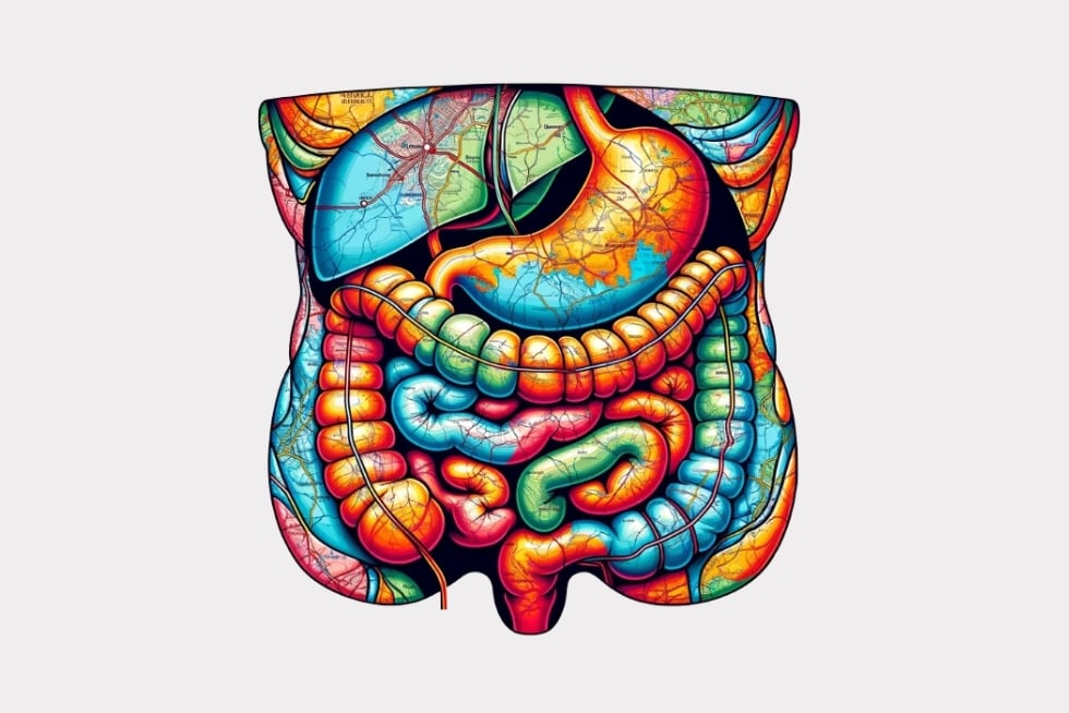
BodyMaps provides a 3D digital atlas of the human body, conceptually similar to Google Maps, but focusing on human anatomy. BodyMaps offers three unique features:
- Semantic segmentation of anatomical structures, such as the cardiovascular system, skeleton, muscles, and gastrointestinal tract.
- Tumor screening across various structures, including the lungs, abdomen, brain, bones, heart, and blood vessels.
- Support of multiple clinical tasks, such as image registration, disease quantification, tumor stage estimation, report generation, etc.
The BodyMaps Project has now contributed a total of 241,336 computed tomography volumes with detailed, per-voxel annotations of 300 anatomical structures and associated tumors. This dataset has already attracted 50+ leading research teams worldwide, driving innovation from classical methods (e.g., U-Net and its variants) to cutting-edge foundation models.
In 2009, before the advent of ImageNet, it was challenging to develop AI algorithms that were robust to different domains using small or even medium amount of labeled data. The same situation presents in medical image analysis today, which is in dire need of its own ImageNet moment—in which a large amount of data is available, high-quality annotations are performed, multiple domains (e.g., hospitals) are covered, and the dataset is attached to a widely recognized challenge.
To this end, BodyMaps presents unprecedented data and annotation scales, with over 72.4 million organ/tumor masks and 81.7 million annotated images that are taken from 145 hospitals across 19 countries and manually inspected by expert radiologists.