In: Machine Learning and Artificial Intelligence

Zongwei Zhou awarded $2.8 million NIH grant
- August 4, 2025
- Machine Learning and Artificial IntelligenceMedical Imaging
The National Institutes of Health awarded Zhou and his team a four-year, $2.8 million R01 grant to develop an AI system to enhance the detection and monitoring of metastasis in colorectal cancer using patients’ CT scans.

Speak and your X-ray will be imaged
- July 29, 2025
- Machine Learning and Artificial IntelligenceMedical ImagingRobotics, Augmented Reality, and Devices
Johns Hopkins researchers present the voice-controlled X-ray imaging system that earned a Best Paper Award at IPCAI 2025.

Malone Center members among recipients of Johns Hopkins Nexus Awards
- July 23, 2025
- Center NewsMachine Learning and Artificial IntelligenceSystems Modeling and Optimization
The Nexus Awards Program supports a diverse range of programming, research, and teaching activities at the Hopkins Bloomberg Center.

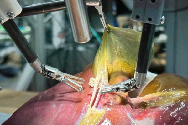
Robot performs first realistic surgery without human help
A system trained on videos of surgeries performs like an expert surgeon.

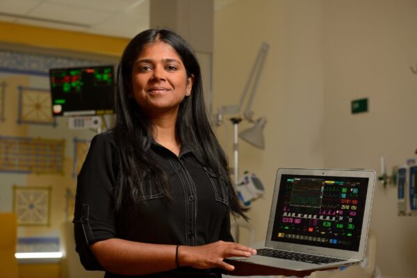
Sepsis detection platform prevents thousands of deaths
- April 25, 2025
- Center NewsMachine Learning and Artificial Intelligence
National Science Foundation funding helped Suchi Saria develop and launch a lifesaving early warning system that uses artificial intelligence to catch sepsis infections before they become deadly.

Malone Center members tackle existential challenges related to assurance, autonomy
- April 17, 2025
- Center NewsMachine Learning and Artificial Intelligence
Selected for the Johns Hopkins Institute for Assured Autonomy’s Challenge Grants, their teams are now completing work that began in 2023.