In: Medical Imaging
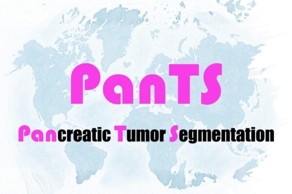
Hold on to your PanTS—there’s a new pancreatic cancer detection dataset in town
- December 10, 2025
- Machine Learning and Artificial IntelligenceMedical Imaging
Developed by a Johns Hopkins-led research team, the Pancreatic Tumor Segmentation Dataset may be the key for training AI models to detect pancreatic cancer early enough to make a difference in patients' survival.
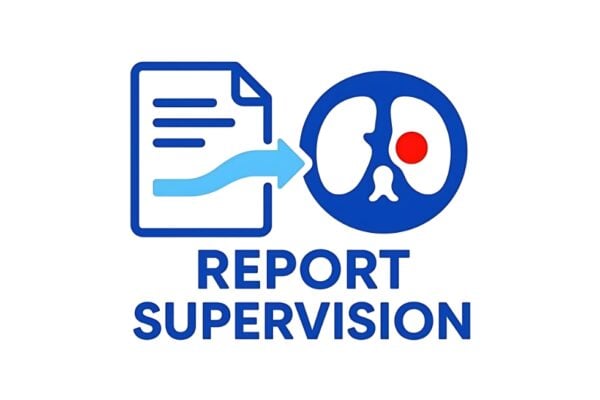
For AI tumor detection, a picture isn’t always worth a thousand words
- September 24, 2025
- Machine Learning and Artificial IntelligenceMedical Imaging
Johns Hopkins researchers have developed a new method that uses existing radiology reports to train AI models to locate tumors on CT scans more quickly and accurately.

Zongwei Zhou awarded $2.8 million NIH grant
- August 4, 2025
- Machine Learning and Artificial IntelligenceMedical Imaging
The National Institutes of Health awarded Zhou and his team a four-year, $2.8 million R01 grant to develop an AI system to enhance the detection and monitoring of metastasis in colorectal cancer using patients’ CT scans.

Speak and your X-ray will be imaged
- July 29, 2025
- Machine Learning and Artificial IntelligenceMedical ImagingRobotics, Augmented Reality, and Devices
Johns Hopkins researchers present the voice-controlled X-ray imaging system that earned a Best Paper Award at IPCAI 2025.
Sharper, safer, more inclusive medical imaging
- April 8, 2025
- Center NewsMedical Imaging
Backed by NIH funding, Muyinatu “Bisi” Bell’s innovative work is enhancing diagnostic accuracy, improving surgical precision, and ensuring equitable health care for all patients.

Muyinatu Bell to develop imaging software for ARPA-H-funded lung cancer project
- November 18, 2024
- Center NewsMedical Imaging
The $13 million award from the Advanced Research Projects Agency for Health will go toward developing a transformative diagnostic tool for lung cancer detection.