In: Medical Imaging

High-speed microscope gives instant look inside living tissue
- February 6, 2026
- Medical Imaging
Developed by Johns Hopkins engineers, the new microscope provides unprecedented, real-time views of living tissue to accelerate patient care and medical research.
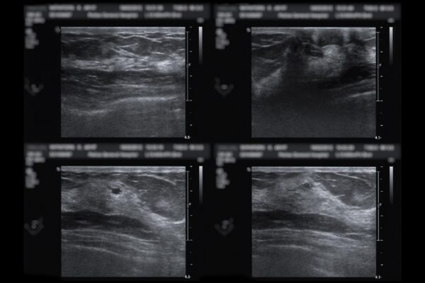
New tech reduces false positives from breast ultrasounds
- January 27, 2026
- Medical Imaging
An advance by Johns Hopkins researchers could spare patients unnecessary follow-up exams and procedures.

Hold on to your PanTS—there’s a new pancreatic cancer detection dataset in town
- December 10, 2025
- Machine Learning and Artificial IntelligenceMedical Imaging
Developed by a Johns Hopkins-led research team, the Pancreatic Tumor Segmentation Dataset may be the key for training AI models to detect pancreatic cancer early enough to make a difference in patients' survival.
For AI tumor detection, a picture isn’t always worth a thousand words
- September 24, 2025
- Machine Learning and Artificial IntelligenceMedical Imaging
Johns Hopkins researchers have developed a new method that uses existing radiology reports to train AI models to locate tumors on CT scans more quickly and accurately.

Zongwei Zhou awarded $2.8 million NIH grant
- August 4, 2025
- Machine Learning and Artificial IntelligenceMedical Imaging
The National Institutes of Health awarded Zhou and his team a four-year, $2.8 million R01 grant to develop an AI system to enhance the detection and monitoring of metastasis in colorectal cancer using patients’ CT scans.

Speak and your X-ray will be imaged
- July 29, 2025
- Machine Learning and Artificial IntelligenceMedical ImagingRobotics, Augmented Reality, and Devices
Johns Hopkins researchers present the voice-controlled X-ray imaging system that earned a Best Paper Award at IPCAI 2025.