In: Medical Imaging
Sharper, safer, more inclusive medical imaging
- April 8, 2025
- Center NewsMedical Imaging
Backed by NIH funding, Muyinatu “Bisi” Bell’s innovative work is enhancing diagnostic accuracy, improving surgical precision, and ensuring equitable healthcare for all patients.

Muyinatu Bell to develop imaging software for ARPA-H-funded lung cancer project
- November 18, 2024
- Center NewsMedical Imaging
The $13 million award from the Advanced Research Projects Agency for Health will go toward developing a transformative diagnostic tool for lung cancer detection.

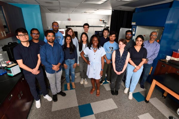
Malone researchers poised to create transformative technologies
Malone researchers are part of two of the four boundary-pushing proposals funded in this year’s cycle of the SURPASS initiative.
New wearable augmented reality device could improve accuracy of spinal tap procedures
- October 3, 2024
- Medical ImagingRobotics, Augmented Reality, and Devices
Johns Hopkins researchers partnered with a local imaging device company to develop an efficient, real-time lumbar puncture guidance system.

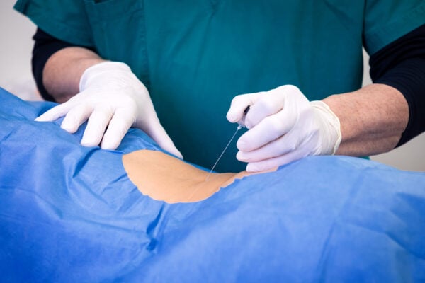
Hopkins team awarded up to $20.9 million in ARPA-H funding to further tumor-removal research
- August 20, 2024
- Center NewsMedical Imaging
A Johns Hopkins-led interinstitutional research team will develop a novel photoacoustic endoscope and fluorescent contrast agent to ensure total tumor removal and preservation of healthy tissue.

Delivering on the promise of personalized medicine
- June 11, 2024
- Center NewsMedical Imaging
Harnessing advances in data science and AI, Whiting School researchers are working closely with clinicians to improve care for a broad array of debilitating conditions.