In: Medical Imaging
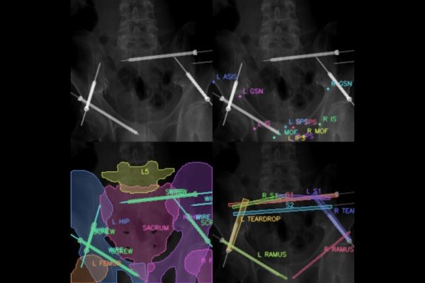
X-ray vision: Tech could improve efficiency of pelvic fracture surgery
- November 22, 2023
- Machine Learning and Artificial IntelligenceMedical Imaging
Johns Hopkins researchers harness the power of machine learning to develop a first approach to X-ray-guided surgical phase recognition.

Medical imaging fails dark skin. Researchers fixed it.
- October 11, 2023
- Medical Imaging
A Johns Hopkins University-led team found a way to deliver clear pictures of anyone's internal anatomy, no matter their skin tone.
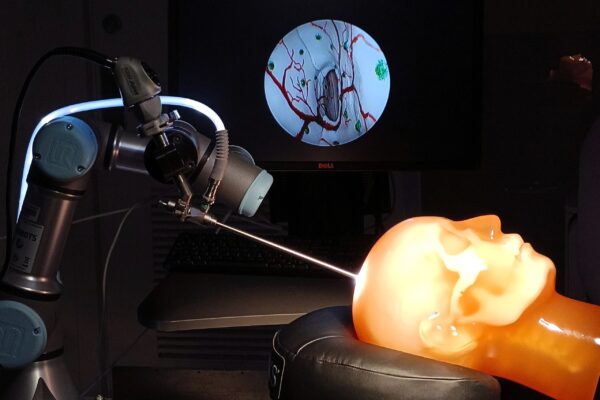
Navigational technology used in self-driving cars aids brain surgery visualization
- August 31, 2023
- Medical Imaging
Johns Hopkins researchers demonstrate the promise of “augmented endoscopy,” a real-time neurosurgical guidance method that uses advanced computer vision techniques.

Muyinatu Bell awarded $1.5 million NIH grant
- August 22, 2023
- Medical Imaging
John C. Malone Associate Professor of Electrical and Computer Engineering Muyinatu A. Lediju Bell has received a four-year, $1.5 million R01 grant from the National Institutes of Health to develop new technology for photoacoustic-guided hysterectomies in an effort to make the procedures safer via informative, real-time feedback.

Photoacoustic imaging gets flexible
- July 31, 2023
- Medical Imaging
In an effort to improve real-time surgical visualization and reduce potential patient discomfort and injury, Johns Hopkins researchers have verified the accuracy and applicability of a minimally invasive live medical imaging method in a new study.

Malone faculty win DELTA Award
Their winning proposal will receive up to $75,000 to help innovate learning through technology.