By: Jaimie Patterson
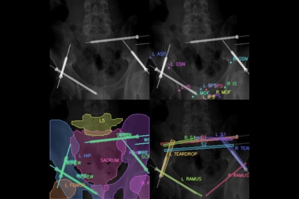
X-ray vision: Tech could improve efficiency of pelvic fracture surgery
- November 22, 2023
- Machine Learning and Artificial IntelligenceMedical Imaging
Johns Hopkins researchers harness the power of machine learning to develop a first approach to X-ray-guided surgical phase recognition.
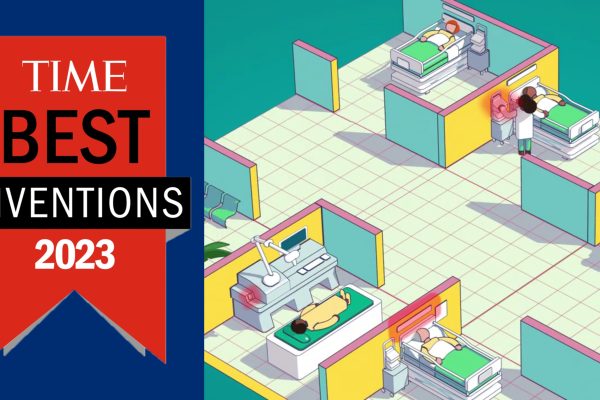
Malone faculty behind one of TIME’s Best Inventions of ’23
- October 30, 2023
- Center NewsMachine Learning and Artificial Intelligence
TREWS predicts patients’ risk of sepsis.

Putting prosthetics research back in touch
- October 25, 2023
- Robotics, Augmented Reality, and DevicesUser-Centered Design
Researchers urge the greater prosthesis engineering community to listen to end users' actual needs.

Johns Hopkins researchers make the case for social media standards on suicide
- October 18, 2023
- Machine Learning and Artificial Intelligence
Johns Hopkins researchers call for the establishment of guidelines that prescribe how modern social media platforms should share pro-social, life-saving education and remove harmful content.

Putting trust to the test
- October 17, 2023
- Machine Learning and Artificial Intelligence
Hopkins researchers unveil new uncertainty quantification methods in an effort to promote appropriate trust in AI use.
Medical imaging fails dark skin. Researchers fixed it.
- October 11, 2023
- Medical Imaging
A Johns Hopkins University-led team found a way to deliver clear pictures of anyone's internal anatomy, no matter their skin tone.