A research team from the Malone Center for Engineering in Healthcare and the Wilmer Eye Institute has received funding from the Retina Society to automate the early detection of sickle cell retinopathy.
Sickle cell disease, the most common inherited blood disorder, can affect every organ system, including the eyes. Over time, proliferative sickle cell retinopathy (PSR) can lead to vision loss and blindness in more severe cases. Thus, it is crucial for clinicians to identify early signs of PSR so they can start treatment faster.
The $50,000 grant, co-sponsored by the International Retina Research Foundation (IRRF), will support the project, “Automated Detection of Proliferative Sickle Cell Retinopathy in Ultra-Widefield Fundus Images.”

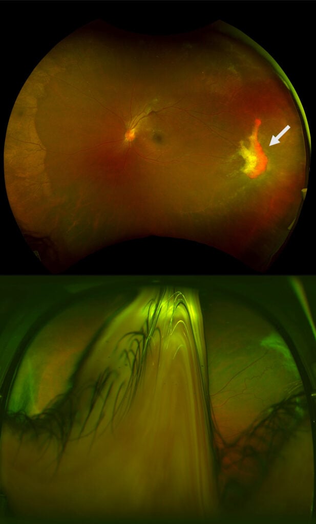
Two ultra-widefield retina photographs in separate patients with sickle cell disease; the top photo detects a sea fan neovascularization complex (white arrow). In the bottom photo, retinal details are obscured by significant eyelid artifact as the patient blinks.
“Through this research we aim to provide a framework that can detect artifacts in fundus images, using machine learning algorithms. These images are typically acquired for severe eye diseases like sickle cell retinopathy at their early stages,” said Craig Jones, an assistant research professor of computer science.
In retinal images of a patient with sickle cell disease, PSR is characterized by a retinal lesion known as a sea fan neovascularization complex. The Hopkins team is working to develop an automated algorithm for detecting sea fan neovascularization from ultra-widefield color fundus photographs. The main goals of the project are to improve diagnostic accuracy of fundus imaging and identify which patients are more likely to experience PSR progression and vision loss.
The research team includes Jones and Mathias Unberath, assistant professor of computer science; and Adrienne Scott, associate professor of ophthalmology at the Wilmer Eye Institute.
